

Advanced Imaging & Surface Analysis
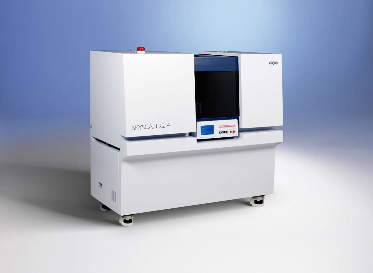
The SkyScan 2214 is an advanced multiscale X-ray Nano-CT system designed for non-destructive three-dimensional imaging of internal structures at submicron and nanoscale resolution. It enables detailed visualization and quantitative analysis of materials, biological tissues, dental specimens, composites, batteries, and engineered components.
Key Features
True 3D spatial resolution below 500 nm
Minimum voxel size of 60 nm
X-ray source range: 20–160 kV
Large sample capacity (up to 300 mm diameter)
Supports multiple detectors for flexible imaging
Non-destructive internal structure analysis
GPU-accelerated reconstruction and advanced 3D visualization software
In-situ testing capabilities (heating, cooling, compression, tensile testing).
Applications
Dental and biomaterial research
Bone and tissue characterization
Additive manufacturing and 3D printing inspection
Composite and fiber analysis
Battery and fuel cell evaluation
Geological and petrochemical studies
Electronics failure analysis
Forensic and paleontological investigations.
Product Overview
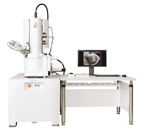
The JEOL JSM-IT800 SHL is a state-of-the-art Field Emission Scanning Electron Microscope (FE-SEM) designed for ultra-high-resolution imaging and microstructural characterization of a wide range of materials. Equipped with advanced field emission technology, the system delivers exceptional surface detail, nanoscale imaging, and analytical capabilities, making it ideal for materials science, biomaterials, nanotechnology, and life science research.
Key Features
High-resolution field emission electron source
Ultra-high magnification imaging
Excellent low-voltage imaging performance
Large specimen chamber for versatile sample accommodation
Automated operation and intelligent navigation
High-depth-of-field imaging
Fast image acquisition and processing
User-friendly graphical interface
Compatible with EDS elemental analysis systems
Advanced imaging modes for detailed surface characterization
Applications
Surface morphology and microstructure analysis
Dental and biomaterials research
Nanomaterials characterization
Failure and fracture analysis
Thin film and coating evaluation
Particle size and shape analysis
Corrosion and wear studies
Semiconductor and electronic materials research
Biological specimen imaging
Additive manufacturing and advanced materials characterization
Technical Capabilities
Nanoscale surface imaging
High-resolution topographical analysis
Elemental composition analysis (with EDS)
Microstructural characterization
Cross-sectional imaging
Particle and pore analysis
Quantitative image analysis
High-vacuum and low-vacuum observation modes
Product Overview
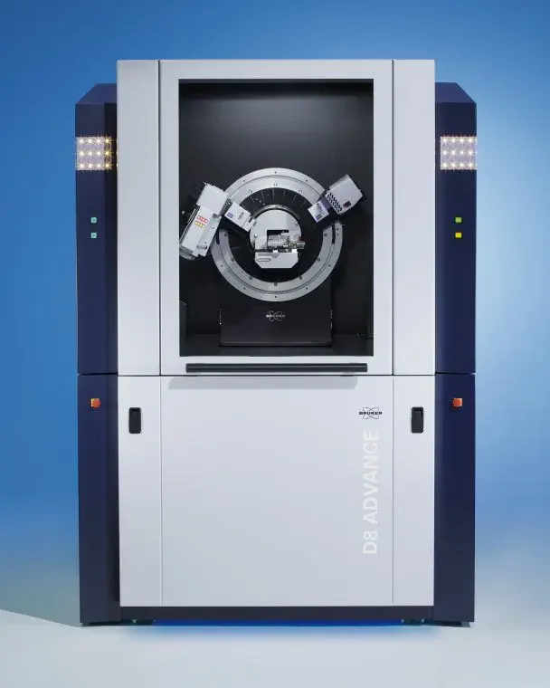
The D8 ADVANCE is a high-performance X-ray diffraction (XRD) system designed for phase identification, quantitative phase analysis, crystal structure determination, residual stress analysis, texture analysis, thin-film characterization, and small-angle X-ray scattering (SAXS). Its modular architecture enables analysis of powders, bulk materials, thin films, fibers, and liquids on a single platform.
Key Features
Advanced X-ray Powder Diffraction (XRD) capabilities
Supports XRD, SAXS, WAXS, and PDF analysis
Automated optics and detector configuration (DAVINCI.DESIGN)
High-precision D8 goniometer with excellent angular accuracy
Dynamic Beam Optimization (DBO) for enhanced data quality
Compatible with multiple detectors, including LYNXEYE XE-T
Automated sample changers for high-throughput analysis
Non-ambient measurements with temperature, humidity, and pressure control options
User-friendly DIFFRAC.SUITE software platform for acquisition and analysis.
Applications
Crystal structure analysis
Phase identification and quantification
Residual stress measurement
Texture analysis
Thin-film characterization
Nanomaterials and advanced materials research
Pharmaceutical analysis
Cement and building materials characterization
Metals and alloys research
Battery and energy-storage materials evaluation
Technical Capabilities
Angular range: <1° to >150° (2θ)
Supports multiple X-ray wavelengths (Cr, Co, Cu, Mo, Ag)
High-speed data acquisition with advanced detectors
Automated switching between diffraction geometries
Suitable for powders, thin films, bulk solids, fibers, and coatings
Product Overview
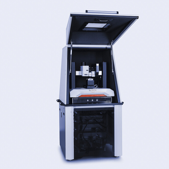
The Anton Paar STEP 700 is an advanced nanomechanical characterization platform that combines Atomic Force Microscopy (AFM) with ultra-nanoindentation and mechanical property mapping capabilities. Integrated with the MCT³, UNHT³, and Nanosurf Nanite AFM modules, the system enables comprehensive analysis of surface topography, roughness, hardness, elastic modulus, adhesion, and nanomechanical behavior at micro- and nanometer scales.
This versatile platform is particularly valuable for biomaterials, dental materials, polymers, coatings, thin films, and advanced material research, providing both morphological and mechanical characterization within a single integrated system.
Key Features
High-resolution Atomic Force Microscopy (AFM)
Three-dimensional nanoscale surface imaging
Ultra Nano Hardness Testing (UNHT³)
Quantitative nanomechanical property mapping
Surface roughness and texture analysis
Elastic modulus and hardness determination
Adhesion and force spectroscopy measurements
High-precision indentation and scratch testing
Non-destructive surface characterization
Automated data acquisition and analysis
Applications
Dental material characterization
Biomaterials and tissue engineering research
Surface roughness evaluation
Nanoindentation and hardness testing
Thin film and coating analysis
Polymer characterization
Nanotechnology and nanomaterials research
Wear and tribological studies
Surface modification assessment
Microstructural and mechanical property evaluation
Technical Capabilities
Nanoscale 3D topographical imaging
Surface roughness measurements (Ra, Rq, Rz)
Ultra-low load nanoindentation
Hardness and elastic modulus determination
Mechanical property mapping
Force-distance curve analysis
Scratch resistance evaluation
Adhesion and deformation studies
Quantitative nanomechanical characterization
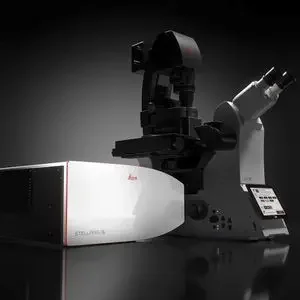
Product Overview
The Leica STELLARIS i5 is a next-generation confocal laser scanning microscope designed for high-resolution fluorescence imaging, live-cell imaging, 3D visualization, and quantitative biological analysis. The system combines spectral detection, photon-counting technology, super-resolution imaging, and fluorescence lifetime-based analysis to provide exceptional image quality and scientific insights.
Key Features
Advanced confocal laser scanning microscopy (CLSM)
High-sensitivity Power HyD detectors with photon-counting capability
Spectral detection for multiplex fluorescence imaging
White Light Laser (WLL) excitation technology
LIGHTNING super-resolution imaging
TauSense fluorescence lifetime imaging tools
Motorized XY stage with automated image acquisition
3D and time-lapse imaging capabilities
Live-cell imaging support with environmental control options
LAS X software for image acquisition, analysis, and visualization
Applications
Cell and tissue imaging
Dental biomaterial characterization
Biofilm and microbial analysis
Surface morphology evaluation
Biomaterials and tissue engineering research
Fluorescence localization studies
Live-cell and developmental biology research
Protein interaction and molecular imaging
Pharmaceutical and biomedical research
Technical Capabilities
Multi-channel fluorescence imaging
Simultaneous detection of multiple fluorophores
Spectral unmixing of overlapping fluorescent signals
Super-resolution imaging beyond conventional confocal limits
Fluorescence Lifetime Imaging (FLIM) support
3D reconstruction and volumetric analysis
Extended spectral detection range up to 850 nm
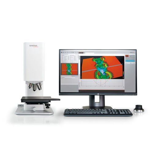
Product Overview
The Sensofar S neox 5-Axis is a state-of-the-art non-contact optical profilometer designed for high-precision surface metrology and three-dimensional surface characterization. Utilizing advanced optical technologies, including confocal microscopy, interferometry, and focus variation, the system provides accurate topographical measurements across micro- and nanoscales. Its unique 5-axis motorized stage enables the analysis of complex geometries and difficult-to-access surfaces with exceptional accuracy.
The S neox is widely used in materials science, dentistry, biomaterials, microelectronics, precision engineering, and industrial quality control for quantitative surface analysis and defect evaluation.
Key Features
Non-contact 3D surface measurement
Integrated confocal, interferometry, and focus variation technologies
High-resolution surface topography imaging
Automated 5-axis motorized positioning system
Nanometer-scale vertical measurement resolution
Large-area surface mapping and stitching
Fast data acquisition and analysis
Quantitative roughness and texture evaluation
Suitable for transparent, reflective, and rough surfaces
Advanced surface metrology software
Applications
Surface roughness analysis
Dental material and restorative material evaluation
Biomaterials characterization
Coating and thin-film assessment
Wear and tribological studies
Microelectronics and semiconductor inspection
Precision manufacturing quality control
Corrosion and degradation analysis
Additive manufacturing research
Surface defect and texture analysis
Technical Capabilities
Three-dimensional surface reconstruction
Surface roughness measurements (Ra, Rq, Rz, Sa, Sq)
Step-height and thickness measurements
Volume and area calculations
Surface waviness and texture analysis
Microstructure and defect evaluation
Large-area stitching and mapping
Quantitative metrology compliant with international standards

Thermal analysis
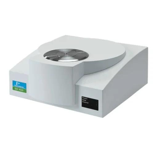
Product Overview
The PerkinElmer DSC 4000 is a high-performance Differential Scanning Calorimeter designed to measure heat flow associated with material transitions as a function of temperature and time. The instrument provides precise analysis of melting, crystallization, glass transition, curing behavior, oxidation, and thermal stability, making it an indispensable tool for material characterization, research, and quality control.
The DSC 4000 is widely used in polymer science, biomaterials, pharmaceuticals, dental materials, nanotechnology, and advanced materials research to understand the thermal properties and phase transitions of materials.
Key Features
High-sensitivity heat flow measurement
Excellent temperature accuracy and reproducibility
Rapid heating and cooling capabilities
Wide operating temperature range
Automated data acquisition and analysis
Precise detection of thermal transitions
Controlled atmosphere operation
User-friendly thermal analysis software
Reliable and reproducible results
Suitable for research and industrial applications
Applications
Glass transition temperature (Tg) determination
Melting and crystallization studies
Polymer characterization
Biomaterial and dental material analysis
Curing and crosslinking behavior evaluation
Phase transition studies
Pharmaceutical thermal characterization
Thermal stability assessment
Nanomaterial research
Quality control and product development
Technical Capabilities
Heat flow measurement as a function of temperature and time
Glass transition (Tg) analysis
Melting temperature (Tm) determination
Crystallization behavior evaluation
Enthalpy measurement
Cure kinetics analysis
Oxidation induction studies
Thermal event characterization
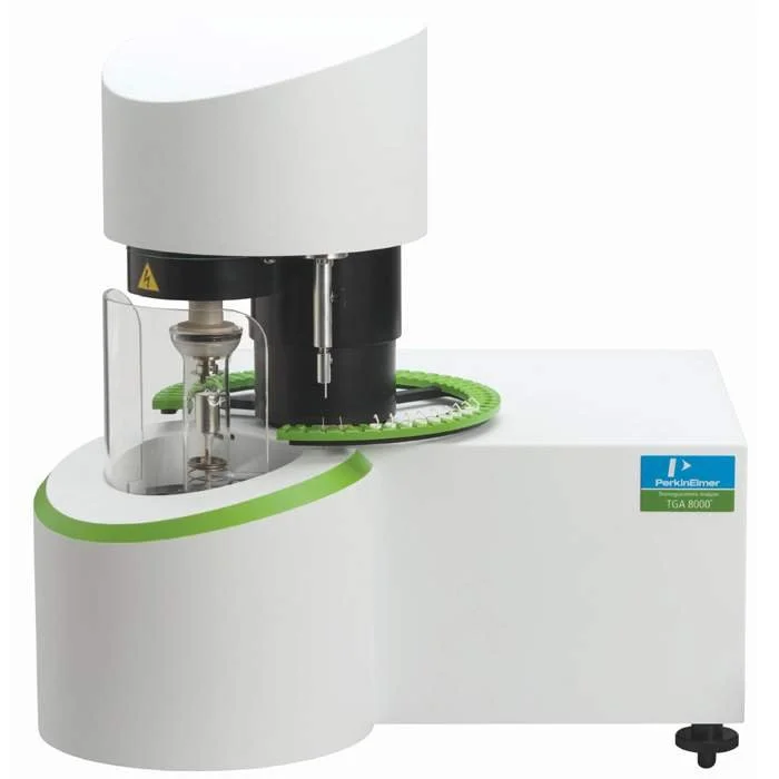
Product Overview
The PerkinElmer TGA 8000 is a high-performance Thermogravimetric Analyzer designed to measure changes in material weight as a function of temperature and time under controlled atmospheric conditions. The system provides precise information on thermal stability, composition, decomposition behavior, moisture content, oxidation resistance, and filler content, making it an essential tool for advanced materials characterization and quality control.
The TGA 8000 is widely used in materials science, polymers, pharmaceuticals, biomaterials, nanomaterials, and industrial research to evaluate the thermal behavior and compositional properties of a wide range of materials.
Key Features
High-sensitivity thermogravimetric measurements
Excellent temperature accuracy and stability
Automated sample analysis and data acquisition
Controlled inert and reactive gas environments
Wide temperature operating range
Precise mass-loss and decomposition analysis
High-resolution thermal stability evaluation
Advanced software for thermal data interpretation
Reliable and reproducible results
Suitable for research and quality control applications
Applications
Thermal stability assessment
Material decomposition studies
Moisture and volatile content determination
Polymer and composite characterization
Biomaterial and dental material research
Filler and ash content analysis
Oxidation and degradation studies
Pharmaceutical thermal analysis
Nanomaterial characterization
Quality control and product development
Product Overview
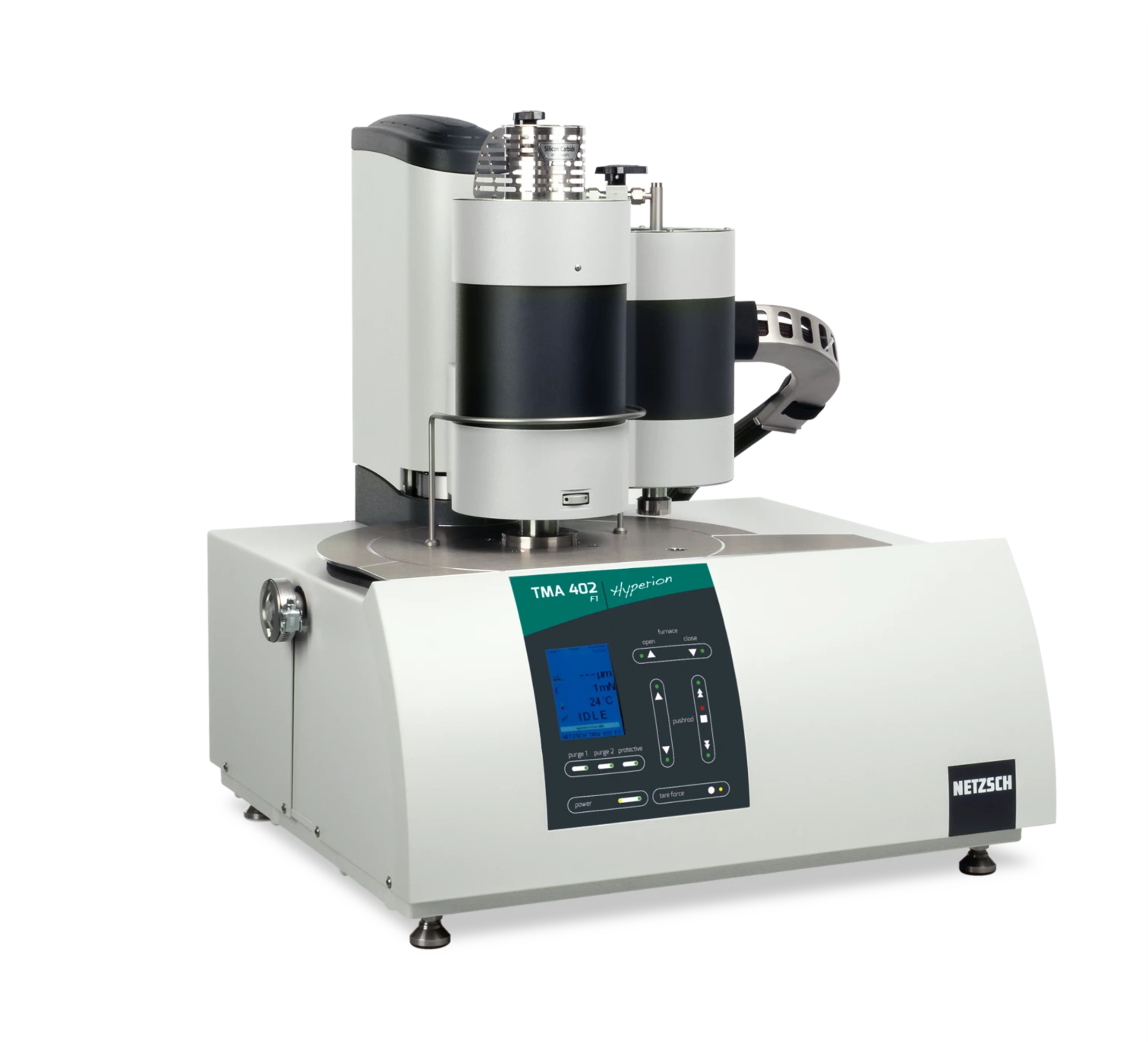
The NETZSCH TMA 402 F3 Hyperion® is a high-precision Thermomechanical Analyzer designed to measure dimensional changes of materials as a function of temperature, time, and applied force. The system provides accurate characterization of thermal expansion, shrinkage, softening behavior, glass transition, and viscoelastic properties, making it an essential instrument for advanced materials research and quality control.
The TMA 402 F3 Hyperion® is widely used for the characterization of polymers, composites, ceramics, metals, biomaterials, dental materials, and electronic components, helping researchers understand material behavior under thermal and mechanical loads.
Key Features
High-precision thermomechanical measurements
Wide temperature operating range
Multiple measurement modes for diverse applications
Automated force control and displacement measurement
High-resolution expansion and contraction analysis
Sensitive detection of glass transition temperatures
Controlled atmosphere operation
Excellent temperature accuracy and reproducibility
User-friendly Proteus® software for data acquisition and analysis
Suitable for research and industrial quality control
Applications
Coefficient of Thermal Expansion (CTE) determination
Dimensional stability studies
Glass transition temperature (Tg) analysis
Softening and deformation behavior evaluation
Polymer and composite characterization
Dental and biomaterial research
Ceramic and metal analysis
Thin film and coating evaluation
Electronic packaging material testing
Quality control and product development
Technical Capabilities
Thermal expansion and contraction measurements
Coefficient of Thermal Expansion (CTE) determination
Penetration and softening point analysis
Dilatometric measurements
Shrinkage and swelling studies
Glass transition detection
Viscoelastic property evaluation
Creep and stress-relaxation investigations
Featured Products
The Atlas Project
A bold reimagining of a timeless brand.


The Echo Project
A bold reimagining of a timeless brand.
Dentin Grinder

A dentin grinder refers to a unique tool utilized in grinding the extracted teeth into dentin useful in autogenous grafts. The gold standard of bone grafting material is autogenous grafts since the patient body generates them

Leica SP1600 saw microtome

Cost of using the facility: Starts from INR 300
The Leica SP1600 saw microtome is specially designed for the sectioning of hard tissues / materials without destroying the morphology of the specimens for various testing and investigation purposes. Section thickness of approximately 50 microns can be achieved under optimal conditions.
The Leica SP1600 saw microtome is specially designed for the cutting of extremely hard and brittle industrial materials embedded in methylmetacrylate with a maximum size of 35 mm in diameter.
Bainpol VT (Single Disc Table Top Grinder/Polisher)

Grit Ranges from 200 to 1200 for various levels of grinding
Quick change disc design enables discs with different grits / polish cloth to be used in desired steps.
DIP coater with IR dryer
The presence of an Infra-Red dryer in it. Inside the unit there is an infrared heater, which offers a maximum temperature of 200°C from ambient. After each dip, the Infrared heater helps in drying the substrate. It provides uniform heating to the substrate. The temperature is swiftly attained so that the time taken for the dipping process is greatly reduced. The coating thickness can be easily controlled by adjusting the withdrawal rate and the viscosity of the coating solution. Another advantage of this model is that it minimizes the energy consumption as the heater will be activated only after detecting the substrate and hence it can provide the substrate with accurate temperature.
Dip coating applications include:
Multilayer sensor coatings
Implant functionalist
Hydro gels
Sol-Gel nano particle coatings
Self-assembled mono layers
Layer-by-layer nano particle assemblies.

Surface profilometer SJ 310

Stylus profilometers use a probe to detect the surface, physically moving a probe along the surface in order to acquire the surface height. This is done mechanically with a feedback loop that monitors the force from the sample pushing up against the probe as it scans along the surface. A diamond stylus is moved vertically in contact with a sample and then moved laterally across the sample for a specified distance and specified contact force. A profilometer can measure small surface variations in vertical stylus displacement as a function of position. A typical profilometer can measure small vertical features ranging in height from 10 nanometres to 1 millimetre. The height position of the diamond stylus generates an analog signal which is converted into a digital signal, stored, analyzed, and displayed. The radius of diamond stylus ranges from 20 nanometres to 50 μm, and the horizontal resolution is controlled by the scan speed and data signal sampling rate. The stylus tracking force can range from less than 1 to 50 milligrams.
Digital Gloss Meter

Gloss Unit : 0 to 1000
Measuring Time 0.5 s
Bioluminometer
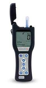
The Bioluminometer allows to instantly verify the microbial contamination of a surface or a liquid, with a bioluminescent reaction.It "read" a light signal that is an output from a sampling device that was passed on the contaminated surface or in the water to be analyzed. During the sampling, the sampling device picks up a cellular molecule, I'ATP (adenosine triphosphate) which is present in all living organisms. When I'ATP is placed in contact with the reaction mixture present in the sampling tube (containing luciferin and luciferase), a reaction takes place that leads to the production of light (photons). This light is measured by the luminometer and converted into a numerical value shown on the display. Higher the microbial contamination is, higher the amount of ATP collection was and, consequently, higher the light produced and the value measured by luminometer would be higher.
Spectrophotometer CM-5
These high quality portable spectrophotometers measure light reflected from a given surface or object. Konica Minolta's portable handheld spectrophotometers can be used to measure solid, opaque, clear, or even translucent samples.

VITA EASY SHADE ADVANCE 4.0
Flow cytometry is a technique that allows characterizing and differentiating between multiple discrete populations of cells based on their light scatter and fluorescence properties as they flow in a single cell file across a tightly focused light beam. Over the last two decades, in adition to being widely used in research settings, it has increasingly become an indispensable tool for screening, diagnosing, prognosticating or monitoring treatment in a wide spectrum of clinical settings.
Our Clinical Flow Cytometry Facility is a state-of-the-art facility utilizing up to 12-color flow cytometric analysis. Our facility is equipped with BD FACSLyric™ Flow Cytometer
NexION® 1000 ICP-MS

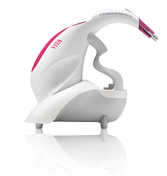
Vita Easy Shade Advance is a digital spectrophotometer to analyse shade and colour stability of aesthetic restorative materials. With calibration block holder G Power supply with universal adapter kit, USB Bluetooth module.
ADVANCED IMMUNOHAEMATOLOGY AND FLOW CYTOMETRY

PerkinElmerSCIEX, inductively coupled plasma mass spectrometry (ICP-MS) has become the fastest growing trace-element detection technique covering a diverse range of applications. Hurdles in successfully coupling an ICP source to a mass spectrometer were initially overcome with design breakthroughs such as a center-tapped ground coil to minimize plasma potential and eliminate secondary discharge between the plasma and the grounded MS interface. Further evolution of plasma RF generators led to a unique free-running design where impedance changes in the plasma were matched instantaneously through small changes in the frequency with no moving parts. In addition, plasma potential was kept at minimum by electronically balancing the plasma with respect to the ground potential.

A densitometer is used to accurately measure the photographic (optical) density at any spot on a radiographic film. For most types of densitometers the size of the measured area is approx. 1 mm2 . The measuring range runs from density 0 to 4
Densitometer, device that measures the density, or the degree of darkening, of a photographic film or plate by recording photometrically its transparency (fraction of incident light transmitted).

Malvern Zetasizer
Particle size: Zetasizers can measure the size of particles from sub-nanometers to several micrometers.
Zeta potential: Zetasizers can measure the charge of particles in a liquid.
A Zetasizer is a particle size analyzer that uses light scattering to measure the size, charge, and molecular weight of particles in a liquid. Zetasizers are used in many industries, including food and beverage, pharmaceuticals, and nanomaterials.
Dynamic Light Scattering (DLS): Measures particle size
Electrophoretic Light Scattering (ELS): Measures particle charge and mobility